
Professional and painless root canal treatment – save your tooth from extraction!
Microscopic endodontics is the key to successful dental treatment.
No pain, no stress – just a healthy smile that lasts.
Join our satisfied patients.
Book a consultation today!
Root canal treatment – Endodontics Szczecin
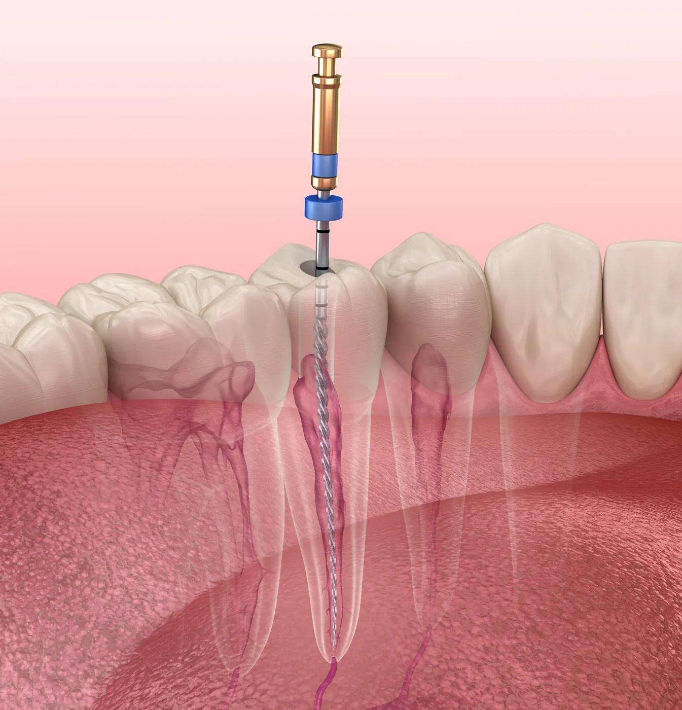
Root canal treatment, also known as endodontics, is an advanced dental procedure aimed at saving a tooth from extraction by removing infected or damaged tissue from inside the tooth, followed by thorough cleaning and sealing of the root canals. At our clinic, we place great emphasis on techniques that effectively restore the function of the patient’s natural teeth.
With the use of state-of-the-art diagnostic equipment, a specialised microscope and precise canal-filling techniques, we offer nearly unlimited treatment possibilities. Thanks to modern technologies and methods, root canal treatment is painless and, in many cases, can be completed in a single visit, significantly reducing treatment time.
Choose professional endodontic care to maintain your oral health and prevent more serious issues in the future.
When is a Root Canal Treatment Necessary?
The primary indication for a root canal treatment is irreversible inflammation of the dental pulp (pulpitis), most commonly caused by untreated tooth decay. This is why regular dental check-ups are so important. Routine visits allow for early detection of dental issues and enable less invasive treatments.
Common symptoms of advanced pulp inflammation include severe, throbbing tooth pain, which often leads patients to seek urgent dental care. Inflammation in the affected area may also cause redness and swelling, and in some cases the patient may develop fever.
Treatments:
Primary Root Canal Treatment
This is an effective method for treating teeth with advanced inflammation and is performed when irreversible pulpitis has occurred. The procedure involves removing the diseased tissue from inside the tooth and root canals. After thorough cleaning, widening, and disinfecting, the canals are tightly sealed with a specialised filling material. Depending on the extent of tooth damage, the crown may be restored with a composite filling or reinforced with a post and core, followed by a prosthetic crown.
To assess the severity of inflammation, an X-ray is taken before the procedure. During treatment, additional X-rays are performed to ensure the canals have been properly cleaned and filled.
Secondary Root Canal Treatment (Re-Endodontics)
This treatment is necessary when inflammation develops or persists in a tooth that has previously undergone endodontic therapy. This condition, often referred to as periapical disease, involves infection and inflammation of the bone tissue surrounding the tooth root. Over time, this infection may progress and form a cyst.
This treatment is necessary when inflammation develops or persists in a tooth that has previously undergone endodontic therapy. This condition, often referred to as periapical disease, involves infection and inflammation of the bone tissue surrounding the tooth root. Over time, this infection may progress and form a cyst.
Common causes of complications after a root canal treatment include:
- Bacteria remaining inside the canal or periapical area
- Undetected additional canals during the initial treatment
- Canals that were not completely treated due to limited access or technological limitations (in our clinic, we use a specialised Stomatol microscope, which enables effective treatment of even the most challenging root canals)
- A broken endodontic instrument remaining in the root canal
- New deep tooth decay
- Infection spreading from neighbouring roots
- Fractured or cracked tooth crown
Complex Root Canal Treatments:
- Removal of Broken Endodontic Instruments – During the cleaning of root canals, particularly curved or obstructed ones, a fragment of an instrument may break and become lodged inside the canal. The presence of a broken instrument prevents proper cleaning, disinfecting, and sealing of the canal, allowing bacteria to remain and potentially leading to treatment failure. Removing such fragments requires advanced skills and specialised equipment, including a surgical microscope and an ultrasonic device with dedicated tips. Our clinic is equipped with state-of-the-art technology, including a Swiss Kaps microscope, which allows for highly magnified procedures, and an EMS ultrasonic device for effective instrument retrieval.
- Clearing Blocked (Obliterated) Root Canals – Successful endodontic treatment requires access to the full length of the root canals. However, obliterated (blocked) canals pose a significant challenge and often require microscopic assistance. The most common causes of canal blockage are age-related pulp degeneration or calcification associated with disease. Chronic inflammation can lead to calcium deposition forming inside the canal, gradually narrowing it and making treatment more difficult.
- Sealing Canal Perforations with MTA – Perforation of a root canal can occur due to tooth decay, resorption, or accidental damage during endodontic treatment. It is most common in curved or obstructed canals. Successful perforation repair depends on the size and location of the perforation. When the canal wall is damaged, a dedicated MTA (Mineral Trioxide Aggregate) material is used to seal the area before the final root filling. Owing to the precision required, this procedure is performed under high magnification using a dental microscope.
Treatment Under a Microscope – When Precision Matters!
Advanced Dental Treatment Under High Magnification!
Root canal treatment under a microscope is a highly effective and safe method offered by modern dentistry. Using a microscope, the dentist can perform complex procedures with extreme precision, viewing the treatment area under high magnification. This allows for accurate localisation, thorough cleaning, and precise sealing of root canals. Additionally, it enables the closure of root perforations and the repair of the tooth chamber floor.
Microscopic endodontics is particularly beneficial in cases involving narrow root canals and root apex resections, helping to avoid tooth extraction and preserve the natural tooth.
Microscopic Root Canal Treatment – What You Should Know?
Advantages of Root Canal Treatment Under a Microscope.
This technique offers many important benefits:
- Enhanced visibility of tooth structures under high magnification,
- Maximum precision and accuracy, preventing discomfort and the need for extractions,
- Modern diagnostic and treatment capabilities, especially for teeth with complex anatomy.
Molar teeth often have multiple root canals (four or even five), which of which are difficult to detect with the naked eye. A microscope allows for the precise identification of all canals, which is crucial because undetected canals increase the risk of treatment failure and can prolong pain caused by persistent infection.
Thanks to high magnification, a microscope enables:
- Access to extremely narrow or blocked root canals,
- Removal of broken endodontic instruments from within the canal,
- Removal of old root canal fillings and post-core restorations (retreatment),
- Closure of root perforations.
Microscopic endodontics is a far less invasive and more cost-effective alternative compared to tooth extraction followed by replacement with an implant or bridge.
How Does Root Canal Treatment Under a Microscope Work?
The process is similar to traditional root canal treatment, but with the added advantage of enhanced visibility through the microscope.
With various magnification levels, previously invisible canals, curvatures, and calcifications (hardened deposits blocking the canal) become visible, making treatment easier and more effective.
Is Root Canal Treatment Under a Microscope Painful?
No – the procedure is performed under local anaesthesia, making it completely painless.
How Many Appointments Are Needed for Microscopic Root Canal Treatment?
In most cases, the treatment can be completed in a single visit.
However, if extensive infection or complications are present, multiple sessions may be required.
How Can You Avoid Microscopic Endodontic Treatment?
- Consistent Oral Hygiene – Brush your teeth at least twice a day, use dental floss and fluoride mouthwash to maintain good oral health.
- Healthy Diet – Reduce the consumption of simple carbohydrates (sugary foods and drinks) and replace them with water and healthier alternatives.
- Regular Dental Check-Ups – Visit your dentist at least once a year to detect cavities at an early stage. Preventive care helps avoid deep decay and pulp infections, ultimately reducing the need for complex root canal treatments.
Trust Our Clinic!
With the latest technologies and the experience of our doctors, we are able to successfully treat even the most challenging dental cases.
Modern Technologies – Our clinic is equipped with a dental microscope and ultrasonic devices, allowing for precise root canal treatment, removal of broken instruments, and clearing difficult root canals.
Experienced Doctors – Our specialists, with years of experience, handle complex endodontic cases, ensuring the highest standard of care and effective treatment.
Comfort and Safety – We prioritize patient comfort by offering inhalation sedation and a friendly atmosphere during procedures.
Contact us today and discover how we can assist with your root canal treatment!
Contact Us